|
 |
Females:
- Monodelphic-prodelphic. Telogonic ovary antidromously
reflexed, on right side of intestine,
- Oviduct situated on left side of ovary; uterus well developed
- Vulva a transverse slit with thick cuticular walls, located at 2/3
body length.
- Demanian system oncholaimoid type variant D (cf. Belogurov and
Belogurova 1988). Ductus uterinus anteriorly inconspicuous. Uvette
well-developed, spherical with thick sclerotized wall, situated on right
side of intestine at 80%-85% of body length. Main duct anteriorly
forming one short sac with thick sclerotized wall and filled with
sperms, posteriorly forming two sacs on both sides.
- Terminal pores arranged in single circle surrounding body, 3-4
abd anterior to anus.
- Tail gradually tapering throughout its length, 5.5-6.1abd long.
|
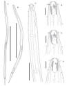
Fotolaimus curvus A. Male body, right lateral view; B. Female
body, right lateral view; C. Male cervical region, right lateral view; D.
Male cephalic region, right lateral view; E. Female cephalic region, right
lateral view; F. Female cephalic region, dorsal view. Scale bars: 1 mm (A,
B); 100 um (C); 20 um (D-F)
Drawings from Shimada et al., 2023 |
|
Males:
- Spicules paired, arcuate, distally acute with subterminal opening,
length 1.3-1.5 cbd.
- Gubernaculum plate-like, distally associated with spicules,
proximally curved,
- No precloacal supplement.
- Circumcloacal setae in single circle,
- Three ventral papillae (Fig. 2L) just anterior to cloacal opening,
in single longitudinal row,
- Diorchic. testes opposed and outstretched, telogonic, on right side
of intestine:
- Tail strongly tapering in anterior 1/5, gradually tapering in next
2/5, and almost cylindrical in posterior 2/5, 3.9-5.7 cbd long.
Ref: Shimada et al., 2023
Reported median body size for this species (Length mm; width micrometers; weight micrograms) - Click:
|