Loa loa
Classification:
- Nematoda
-
Chromadorea
Spiruria - Spirurida
- Filarioidea
- Onchocercidae
-
Loa loa Cobbold, 1964
eye worm
A cutaneous filarial parasite of humans
Biology and Epidemiology
Loa loa - a filarial nematode causing loa loa filariasis primarily in Africa and India.
L. loa ind three other filarial nematodes cause subcutaneous filariasis in humans. The other are Mansonella streptocerca,Onchocerca volvulus (causes river blindness), and Dracunculus medinensis (guinea worm).
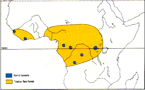
Distribution:
Human loiasis is confined to the rain forest and swamp forest areas of West Africa. It is especially common in Cameroon and on the Ogowe river.
(taken from Peters and Gilles1991)
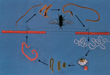
Life Cycle:
In filarial nematodes, Two hosts to complete the life cycle, an intermediate host (often an arthropd) and a primary host (usually a vertebrate). The juvenile stages occur in the intermediate host and the reproductive adult in the definitive (primary) host.
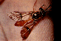
Loiasis is caused by the filarial nematode Loa loa which is transmitted to humans by day-biting Chrysops flies (see picture below taken from Peter and Gilles 1991). Juveniles develop in horseflies, deer flies, yellow flies which infect humans by biting them.- A vector fly bites an infected human host and ingests microfilariae.
- Microfilariae move to the thoracic muscles of the insect host.
- Microfilariae develop into first stage juveniles, second and third stage juveniles.
- Third stage juveniles are the infective stage and travel to the proboscis of fly.
- An infected vector fly bites an uninfected human host; the third stage juvenile penetrates the skin and enters human subcutaneous tissue.
- Juveniles mature into adults, which produce microfilariae that have been found in spinal fluid, urine, peripheral blood, and lungs.
Once inside the human body the infective larvae develop slowly into a mature adult (the process takes about a year). During this period it lives and moves around the fascial layers of the skin. In periods of growth and development Loa loa often makes frequent excursions through the subdermal connective tissues where it is often noticed by the host.
Once they reach maturity the adults mate and
produce sheathed microfilariae 298 x 7.5 μm in size.
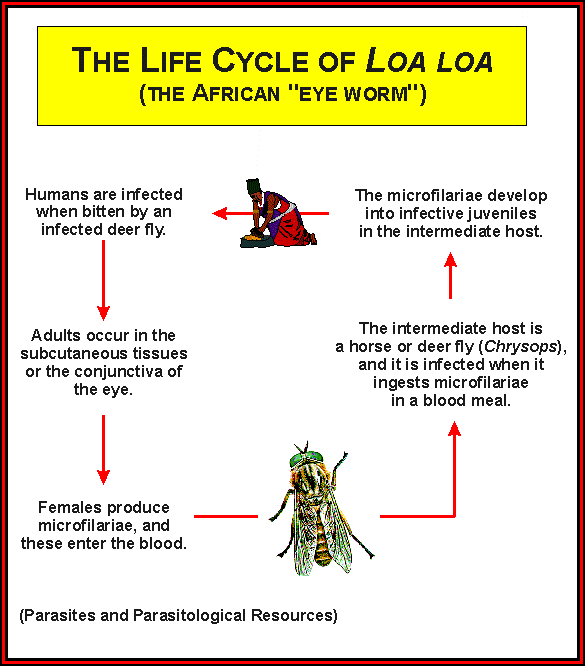
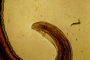
The microfilariae closely resemble the microfilariae of Wuchereria bancrofti however in stained films they assume a stiff angular attitude. The cuticle sheath also does not stain with Giemsa (see picture below taken from Peters and Gilles 1991).
The microfilariae are diurnally periodic in synchrony with their vector and once they reinfect a fly they undergo two stages of growth into infective larvae (in about 10 days time) which can then be transmitted back to humans.
Pathology
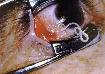
Most of the pathological problems observed in people infected with Loa loa are connected to periods when the migrating adult worms appear near the surface of the skin. The worms often appear around the eye where they can be easily seen and extracted before they damage the conjunctiva (See pictures below taken from Peters and Gilles 1991).
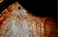

Immune reactions to the migrating worms can also cause calabar swellings in the arms and legs. Recurrent swelling can lead to the formation of cyst like enlargements of the connective tissues around the tendon sheaths. These swellings can be extremely painful when moved. Dying worms can also cause chronic abscesses followed by granulomatous reactions and fibrosis.