Wuchereria bancrofti
Contents
Rev 06/07/2026
Wuchereria bancrofti |
Contents |
Rev 06/07/2026 |
||
|
|
Classification |
|
Hosts | |
|
|
|
Morphology and Anatomy |
|
Life Cycle |
| Return to Wuchereria Menu |
|
Economic Importance |
|
Damage |
|
|
|
Distribution |
|
Management |
| Return to Filariidae Menu |
|
Feeding |
|
References |
| Nemaplex Home Page |
The causal agent of bancroftian filariasis, elephantiasis, lymphatic filariasis
![]()
Two nematode species are associated with elephantiasis, Wuchereria bancrofti and Brugia malayi. They are similar in many aspects of their biology. One primary difference is that brugian filariasis is not as widely distributed as is bancroftian filariasis, although the distributions of the two diseases overlap in many areas.
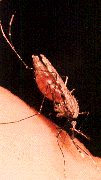
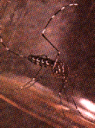
Wuchereria bancrofti is distributed throughout the tropical regions of Asia, Africa, China, the Pacific and isolated locations in the Americas. Current estimates (WHO, 1994) suggest that 100 million people are infected with lymphatic filariae of all types, and most of these cases are bancroftian filariasis. Nocturnally periodic forms occur indigenously in almost every tropical and subtropical country and are very widespread. However they show focal and periodic distribution patterns which are dependent on their vector of transmission. Culcicine (left) or anopheline (center) mosquitoes are the main vectors of the nocturnally periodic forms of W. bancrofti, while day biting Aedes polynesiensis transmit the subperiodic form in various pacific islands (right) (map and pictures taken from Peters and Gilles 1991).

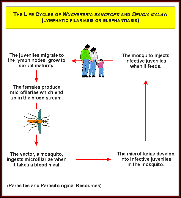
The third-stage infective nematode larvae (L3i) enter the blood through the wound made by the mosquito. They then migrate to the nearest lymph gland where they mature into the thread like adult worms about 3 months to 1 year later. The average incubation time before patency is about 15 months. The mature adults can survive for 5 to 10 years and the damage of the lymphatic vessels they cause and the immune system's response to their presence (and that of microfilaria and newly inoculated L3i) can result in the various symptoms.
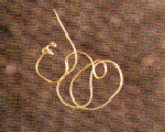
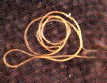
Once male (left) and female (right) nematodes mate, the female viviparously produces microfilariae (first stage larvae or L1) which then move through the circulatory system and collect in arterioles of the lung during the day and emerge at night (if nocturnally periodic) when night biting mosquitoes are most active.
Once the microfilariae have entered an appropriate mosquito host through its blood meal they penetrate the insect's gut wall and move to the thoracic muscles where they mature (through two life stages) into third-stage infective larvae (pictures taken from Peters and Gilles 1991).
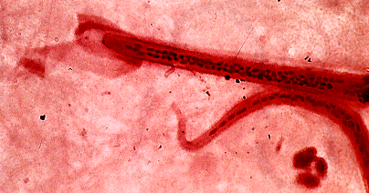
The microfilariae of W. bancrofti can be identified in blood smears by their sheath, size (280 x 7 micrometers), and the anterior V spot near the head and posterior V spot near the tail (picture taken from Peters and Gilles 1991).
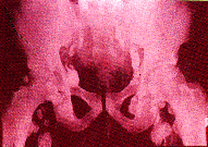
Following infection with third stage larvae there is usually a period of vigorous immune response to the invading larvae. If the larvae are not cleared from the body during this period then the various pathologies associated with filarial infection can develop. Most of these conditions do not appear to arise from the effects of the nematodes themselves but from immune reactions to their presence. The most pronounced of these is the damage to the lymphatic vessels which is mediated by the immune system's response to the adult worms living in them. These immune responses (Lymphangitis) are characterized by inflammation of the affected area (which are usually extremities) and fever. Repeated episodes of lymphangitis lead to the formation of fibrous and calcified tissues (as seen in the X-ray below, picture taken from Peters and Gilles 1991) in and around the lymphatic vessels.
This can then result in gross enlargement of the effected lymph nodes and the pictures below (taken from Peters and Gilles 1991) show two examples of such conditions which are called elephantiasis.
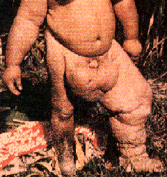

With W. bancrofti infections these enlargements are usually unilateral and the incapacitating deformities often require radical surgery to remove the surplus fibrous and calcified tissues. The microfilariae in the blood and lungs can also cause an IgE mediated allergic response which results in asthma like symptoms. This condition is called tropical eosinophilia and is treatable with macro and microfilaricides