Revised 06/07/26
The nematodes of the family Anisakidae were probably first recognized in fish hosts ine 13th century, in marine mammals in the early 1970s, as an occasional human disease in 1867, and as a more common human infection in the 1950s and 1960s. Between 1970 and 1976, eight human cases of anisakiasis were documented from North America (Myers, 1976).
Knowledge of nematodes responsible for human anisakiasis spans almost 500 years, paralleling advancement of research from visual observation-through development of the microscope, electron microscope (Myers, 1976) and the development of biolochemcal and molecular technology.
Larvae of the Anisakidae are a major problem for commercial fishing industries and are potential human health hazards, both as causative agents of anisakiasis, and as potential food-borne allergens (Nadler et al., 2005).
The following detailed descriptions and lifecycles of nematodes associased with Anisakiasis are provided courtesy of CDC, the Centers for Disease Control and Prevention
Causal Agents
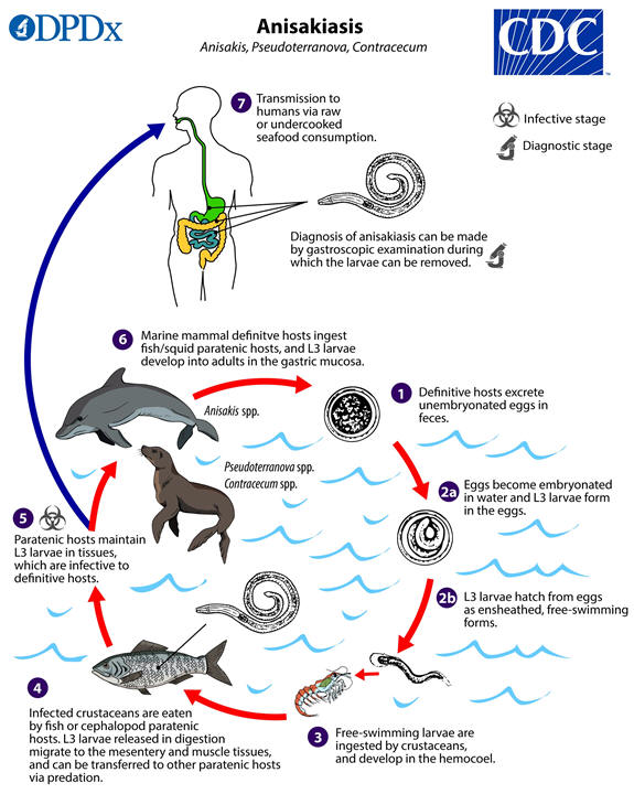
Anisakiasis is caused by the ingestion of larvae of several species of ascaridoid nematodes (roundworms), which are sometimes called “herringworm”, “codworm”, or “sealworm”, in undercooked marine fish. Known human-infecting anisakid species include members of the Anisakis simplex complex [A. simplex sensu stricto, A. pegreffii, A. berlandi (=A. simplex C)], the Pseudoterranova decipiens complex (P. decipiens sensu stricto, P. azarasi, P. cattani, and others), and the Contracecum osculatum complex. Recent genetic studies have revealed high diversity within these anisakid groups, suggesting additional cryptic species are likely represented in zoonotic infections.
Adult stages of anisakid nematodes reside in the stomach of marine mammals,
where they are embedded in the mucosa in clusters. Unembryonated eggs produced
by adult females are passed in the feces of marine mammals  .
The eggs become embryonated in water, undergoing two developmental molts
.
The eggs become embryonated in water, undergoing two developmental molts  ,
and hatch from the eggs as free-swimming ensheathed third-stage (L3) larvae
,
and hatch from the eggs as free-swimming ensheathed third-stage (L3) larvae  .
These free-swimming larvae are then ingested by crustaceans
.
These free-swimming larvae are then ingested by crustaceans  .
The ingested larvae grow within the crustacean hemocoel, and become infective to
fish and cephalopod paratenic hosts. After preying upon infected crustaceans,
the digested L3 larvae migrate from the paratenic host intestine into the
abdominal cavity, and eventually to the tissues of the mesenteries and skeletal
muscle. Through predation, tissue-stage L3 larvae can be transmitted among
paratenic hosts
.
The ingested larvae grow within the crustacean hemocoel, and become infective to
fish and cephalopod paratenic hosts. After preying upon infected crustaceans,
the digested L3 larvae migrate from the paratenic host intestine into the
abdominal cavity, and eventually to the tissues of the mesenteries and skeletal
muscle. Through predation, tissue-stage L3 larvae can be transmitted among
paratenic hosts  .
Fish and squid maintain L3 larvae that are infective to humans and marine
mammals
.
Fish and squid maintain L3 larvae that are infective to humans and marine
mammals  .
.
When fish or squid containing third-stage larvae are ingested by definitive host
marine mammals, the larvae molt twice and develop into adult worms  .
After ingestion by humans, the anisakid larvae penetrate the gastric and
intestinal mucosa, causing the symptoms of anisakiasis
.
After ingestion by humans, the anisakid larvae penetrate the gastric and
intestinal mucosa, causing the symptoms of anisakiasis  .
.
Gibson, D.I. 1983. Systematics of ascaridoid nematodes - a current assessment. In: Concepts in nematode systematics : Proceedings of an international symposium held jointly with the Association of Applied Biologists, in Cambridge / edited by A.R. Stone, H.M. Platt, and L.F. Khalil
Myers, B.J. 1976. Research Then and Now on the Anisakidae Nematodes. Transactions of the American Microscopical Society, 95:137-142.
Nadler, S.A. 1987. Biochemical and Immunological Systematics of Some Ascaridoid Nematodes: Genetic Divergence between Congeners. The Journal of Parasitology, 73:811-816
Nadler, S.A., D'Amelio, S., Dailey, M.D. Paggi, L., Siu, S., Sakinari, J. A. Molecular phylogenetics abnd diagnosis of Anisakis, Pseudoterranova, and Contracaecum from Nothern Pacific marine mammals. J. Parasitol. 91:1413-1429.
Want more information about nematodes? Go to Nemaplex Main Menu.