(Guinea worm)
Rev. 06/07/2026
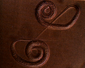
Female 60 cm in length, reported up to 3 m. Male much smaller, 1.2-2.9 cm long. Juveniles (larvae) 500-700 m long.
The
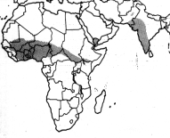
Guinea worm is found predominantly in Africa from Senegal (west) to Ethiopia
(east).
Also found in India, Pakistan, Saudi Arabia, and Yemen.
Inhabits freshwater lakes and rivers.
Dracunculus medinensis, the guinea worm, historically infected approximately 10 million people per year. An eradication program led by the Carter Center has drametically reduced the number of infections and there is hope of eradication of the disease. The majority of human infections occur in parts of West Africa, East Africa, and India.
In south-eastern Nigeria the guinea worm disease, dracunculiasis, is responsible for an 11.6% decrease in the total rice crop, valued at $20 million (how? contamination? labor inefficiencies?).
The guinea worm has been blamed for the majority of school absences in many parts of Africa, wasting valuable time and resources in already-poor third world countries.
Juvenile stages live in fresh water and infect copepods (e.g. Cyclops - water fleas), the secondary host.
The guinea-worm like all filarial nematodes goes through six developmental stages. The however unlike any other filarial parasite that can be transmitted to humans the infective larvae enter the body through the ingestion of various species of the freshwater crustacean Cyclops in contaminated water.
Cyclops sp. (Peters and Gilles 1991).
![]()
Inside the definitive (vertebrate) host, the ingested Cyclops is destroyed by stomach acids. The free larvae penetrate the gut lining and migrate to subcutaneous tissues via the lymphatic system. This process takes approximately 43 days and once in subcutaneous tissue the worms mature slowly, reaching full development in one year.
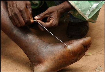
As adults, the nematodes mate. The small male (1.2-2.9 cm long) dies and is absorbed into the larger female (60 cm long). When the embryos in the uterus reach maturity, the female nematode migrates to areas of the body in contact with water (90% move to the feet and legs). Once in these areas the worm penetrates the skin, extrudes its uterus through its mouth, and discharges larvae into the water and dies.
 (photograph
from Peters and Gilles 1991)
(photograph
from Peters and Gilles 1991)
The larvae, which measure between 500 and 700 m, can live for 6 days in clean water and 2 to 3 weeks in muddy water (photograph from Peters and Gilles 1991).
The larvae are ingested by the copepod crustacea, Cyclops, which actively chase them. Once ingested, the larvae mature into their infective stage in approximately 14 days and can then reinfect humans.
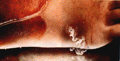
After mating inside the definitive (human) host, the female burrows through the skin, creating an ulcerated area, usually in a lower part of the host's anatomy.
When the ulcerated area is exposed to water (as in a river or lake), the female's uterus ruptures, releasing many juveniles into the water.
Feeding on blood, impairment of lymphatic system(?), skin ulceration.
The female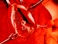 guinea-worm
lives in the connective tissues of the limbs and trunk usually without
noticeable pathological conditions. Although heavy infestations in the
joints can cause arthritic conditions and require the removal of the worms
(picture from Peters and Gilles 1991), most pathology is associated with
infection occurring when the female dies after discharging her larvae.
guinea-worm
lives in the connective tissues of the limbs and trunk usually without
noticeable pathological conditions. Although heavy infestations in the
joints can cause arthritic conditions and require the removal of the worms
(picture from Peters and Gilles 1991), most pathology is associated with
infection occurring when the female dies after discharging her larvae.
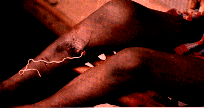 The
death of the worm causes the formation of an abscess which, when secondarily
infected, results in cellulitis and local blistering of the skin (picture from
Peters and Gilles 1991).
The
death of the worm causes the formation of an abscess which, when secondarily
infected, results in cellulitis and local blistering of the skin (picture from
Peters and Gilles 1991).
Also, chills, fever and local painful swellings commonly precede the emergence of the worm. D. medinensis has also been found coiled in the hernial sac and in the placenta, causing bleeding in pregnancy.
There is a worldwide effort to eradicate the Guinea worm. Organizations such as the Task Force for Child Survival and Global 2000 list this as one of their primary objectives.
Spread of the Guinea worm can be prevented by filtering drinking water through a cotton cloth or by treating drinking water with the chemical Abate (which has been tested to be safe for human consumption, and it is not known to harm birds or fish).
The best method for defeating dracunculiasis seems to be supplying villages with safe drinking water.
Material from Mark Potter, 1995 and from the Filarial Genome Network