Photomicrograph by Sung-Jong Hong (The Korean Society for Parasitology).
Rev 05/28/25
Class: Enoplea
Sub-class:
Dorylaimia
Order: Trichinellida
Parasites of vertebrates. All species have at least one life stage that feeds on cells or tissues. Characterised by a stichosome.
Stichosomes occur in two orders of the Nematoda: Trichinellida, with at least six families, and Mermithida, with two families. Recent phylogenetic analysis based on a synthesis of molecular and morphological data suggest that the stichosome may be an example of parallel evolution in the Trichinellida and Mermithida (De Ley and Blaxter, 2002; Ferris, 2007).
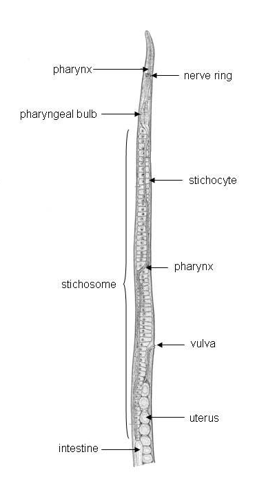
The pharynx is narrow and thin-walled anteriorly and which, posteriorly, is surrounded by unicellular, glandular stichocytes, each with a duct into the pharyngeal lumen.
The pharynx extends one-fourth to nine-tenths of the body length in various
taxa and is almost devoid of musculature. The region of the pharynx surrounded
by stichocytes is
known as the stichosome.
|
|
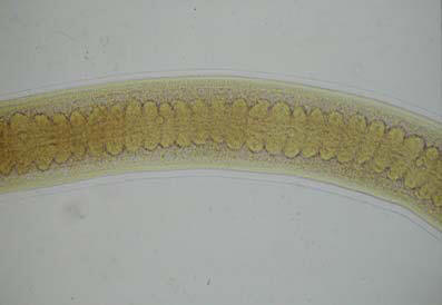
| Figure 1. The stichosome of Trichinella spiralis. Adapted from Chitwood, 1930. | Figure 2. A portion of the stichosome of the whipworm, Trichuris
trichiura. Photomicrograph by Sung-Jong Hong (The Korean Society for Parasitology). |
In Trichinella spiralis, encysted larvae are ingested in infected muscle tissue (raw or undercooked pork is the classic example in the case of trichinosis in humans).
The cyst surrounding the larva is digested in the new host and the larvae
molt to adults, mate, embed in the intestinal epithelium and females produce
eggs which hatch (1000 larvae
per female in 5 days).
The hatched larvae are distributed via the circulatory system and migrate into surrounding cells, which die unless they are striated muscle fibers.
Secretory products of the stichocytes induce DNA endoreduplication and
transformation of the muscle fiber and into a multinucleate nurse cell which
becomes encapsulated by collagen
and supplied with capillaries (Despommier,
1998; Lee, 2002).
The life cycle continues when the muscle is eaten by another animal.
Chitwood, B.G. 1930. The structure of the esophagus in the Trichuroidea. Journal of Parasitology 17:35-42.
De Ley, P. and Blaxter, M. 2002. Systematic position and phylogeny. Pp 1-30 in Lee, D.L. (ed.). The Biology of Nematodes. Taylor and Francis, London and NY. 635p.
Despommier, D.D. 1998. Trichinella and Toxocara. Pp597-607 in Cox, F.E.G.,
Kreier, J.P. and Wakelin, D. Volume 5, Parasitology in Collier, L., Balows, A.
and
Sussman, M. (eds.). Topley and Wilson’s Microbiology and Microbial
Infections. Arnold, London.
Ferris, H. 2007. Stichosomida. McGraw-Hill Encyclopedia of Science & Technology. http://accesscience.com/abstract.aspx?id=757377&referURL=http%3a%2f%2faccesscience.com%2fcontent.aspx%3fid%3d757377.
Lee, D.L. 2002. Life cycles. Pp 61-72 in Lee, D.L. (ed.). The Biology of Nematodes. Taylor and Francis, London and NY. 635p.
Munn, E.A and Munn, P.D. 2002. Feeding and digestion. Pp 211-232 in Lee, D.L. (ed.). The Biology of Nematodes. Taylor and Francis, London and NY. 635p.
Poinar, G.O. and Hess, R. 1977. Romanomermis culicivorax: morphological evidence of transcuticular uptake. Experimental Parasitology 42:27-33.
Poinar, G.O. 1983. The Natural History of Nematodes. Prentice-Hall, Englewood
Cliffs. 323p.