Dioctowittus


Contents


Rev: 12/12/2023

  Classification Biology and Ecology
Morphology and Anatomy Life Cycle
Return to Dioctowittus Menu Ecosystem Functions and Services
Distribution Management
Return to Cystoopsidae Menu Feeding  References
    Go to Nemaplex Main Menu   Go to Dictionary of Terminology

Classification:

Enoplea
    Dorylaimia
        Trichinellida
            Trichinellina
Trichinelloidea

Cystoopsidae


Dioctowittus Chabaud & Le Van Hoa, 1960

    Synonyms:
     
Back to Top

Morphology and Anatomy:

Thread-like esophagus surrounded by stichocytes, intestine transformed into a trophosome, anterior position of the vulva, character of the eggs and site within the snake host conform with characteristtics of the genus Dioctowittus. Bain and Ghadirian (1967) showed that the esophageal masses correspond to a double row of stichocytes.

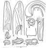
Dioctowittus denisoniae

Fig. 1. Anterior end, male; Fig. 2. Anterior end, female, lateral, with extremely anterior vulva; Fig. 3. Posterior end, male, lateral; Fig. 4. Optical transverse section, male, tail, 0.3mm from posterior end; Fig. 5. Male tail, dorsal; Fig. 6. Posterior end, female; Fig. 7. Egg; Fig. 8. Transverse section, male, c.0.5 mm from ant. end; Fig. 9. Transverse section, male, near origin of trophosome; Fig. 10. Transverse section, male, l mm from tail.

Key: g = testis; t = trophosome; s=stichocyte; v=vas deferens.

Drawings from Jones, 1987

Females:

  • Opisthodelphic, ovary single, apex near posterior end of worm.
  • Single uterus filled with eggs, extendimg length of worm, leading anteriorly into muscular ovejector.
  • Vulva just behind anterior extremity, flush with body surface.
  • Eggs thick-walled, barrel-shaped and markedly flattened. Tuft of fine filaments inserted into mucus plug at each pole, slightly shorter than eggs themselves
  • Eggs cotaining fully-formed larvae.

Ref: Jones, 1987


Males:  

  • Two low ridges on ventral surface of tail, terminating posteriorly short distance from extremity, anteriorly merging into body wall about 2 mm from tail.
  • Monorchic, single testis rounded and reflexed, passing forward to level, or just past level, of commencement of intestine before turning posteriorly.
  • Valvular apparatus at origin of vas deferens; minute genital orifice on posteroventral aspect of tail.
  • Papillae, gubernaculum and spicules absent.

?So, how do sperm get into female?


Body size range for the species of this genus in the database - Click:
Back to Top

Distribution:


Feeding:


Back to Top

Biology and Ecology:

Nematodes of the genus Dioctowittus are highly modified for an existence in the body tissues or body cavity of their hosts. They have a vestigial sensory apparatus and the transformation of the intestine into a trophosome (Bain and Ghadirian 1967).

Back to Top

Life Cycle:


For Ecophysiological Parameters for this genus, click 
 
Back to Top

Ecosystem Functions and Services:

Back to Top

Management:


Back to Top

References:

Bain, O. and Ghadirian, E, 1967. Description d'une nouvelle espece de Dioctowittus (Nematode) et note sur la position systematique du genre. Annales de Parasitologie, 42: 643-650.

Chabaud, A.G. and Le Van Hoa. 1960. Adaptation a la vie tissulaire d'un Nematode Aphasmidien. Comptes Rendus Seances de I'Academie des Sciences, Paris, 251, (17) 1837-1839.

Jones, H.I. 1978. Dioctowittus spp. Chabaud and Le Van Hoa 1960 (Nematoda: Adenophorea) from Australian snakes. J. Helminthology 2:141-145


Copyright © 1999 by Howard Ferris.
Revised: December 12, 2023.